


1. Introduction
Thermal protection technology is critical to the development of hypersonic vehicles, detectors of all types, and missile launchers [1,2]. Specifically, thermal protection systems (TPSs) are based on the selfsacrificing mechanisms of ablative materials through phase changes, chemical reactions, and physical reactions, which consume large amounts of external heat flow and further reduce the continued transfer of heat to the interior by means of the protective materials in the TPS [3,4]. In the early stages of hypersonic vehicle development, the structural design of TPSs became a more prominent topic. For instance, sandwich structures with low thermal conductivity and high-performance benefits were integrated into the structural design of effective TPSs[5], including structures such as corrugated cores [6], multilayer cores[7], foams, and honeycomb cores [8,9]. However, the different material components of these TPSs have different expansion coefficients and exhibit interfacial bonding and other problems [10]. Further, in actual harsh, high-temperature ablation environments, TPS materials are susceptible to erosion by force, heat, and other factors, which affects the overall reliability of the TPS[11,12]. Therefore, a material that combines ablation resistance and excellent thermal insulation properties must be developed.
Although reports have been published on the research and development of anti-insulation integrated materials, such as gradient porous ceramic materials and ablation-resistant phosphate materials [13-16], the thermal conductivity of gradient porous ceramic materials remains a major problem [10]. Phosphate materials were produced in the 1950s by Kingery et al. by using specific phosphoric acid or phosphate blends of ceramic or refractory powders, which were gradually hardened by physico-chemical action [17]. These materials exhibit excellent high temperature resistance, low thermal conductivity, and advantageous mechanical properties [18,19]. The current research on phosphate solutions mainly focuses on aluminum-chromium phosphate solutions [20-22]. In these combinations of aluminum phosphate and chromium phosphate, the introduction of Cr ions reduces the electrostatic effect of the solution system somewhat because the radius of Cr ions is larger than that of Al ions, thus substantially improving the stability of the matrix solution. Most importantly, aluminum-chromium phosphate materials themselves have advantageous properties such as excellent chemical stability, high melting points (1800∘C), low thermal conductivity, and high bonding [23,24]. However, because a temperature resistance of 1800∘C is far from adequate in ultra-high-temperature applications, addressing the temperature resistance of phosphate materials is critical.
Recent work has shown that one of the most effective methods of improving the temperature resistance of phosphate materials is to add high-temperature-resistant oxides [25-27], such as La2O3 [28], MgO [29], and CuO [30]. La-Al phosphate materials [28] obtained by adding an appropriate amount of La2O3 can resist ablation at 2000∘C because of the formation of dense, high-temperature-resistant LaPO4 and AlPO4 layers on the surface after ablation. Zhan et al. [29] prepared an (AlCrMg)x(PO4)y/MgO material with both high-temperature resistance and thermal insulation properties by utilizing MgO and aluminum-chromium phosphate. This material benefited from the formation of a dense protective layer formed by spinel MgCrO2 and MgO under hightemperature ablation and exhibited cooling by 2270∘C over a distance of 15 mm in the thickness direction after 60 s of ablation at 2400∘C. Different high-melting-point oxides have different characteristics. In the study of ultra-high-temperature ceramics, Zr-based ceramics show notable ablation resistance characteristics [30-32], mainly achieved with the help of carbon-nitrogen-zirconium compounds, at high temperatures. A dense ablation-resistant ZrO2 barrier, Zr-O bond energy of 339 kJ, and melting point of up to 2700∘C can be realized, which can block high temperatures from the internal material, thus preventing the continuation of destruction through ablation [33,34]. However, ultra-high-temperature ceramic materials themselves have high thermal conductivity, which affects their use in thermal insulation applications [10], and the direct bonding of mixed ZrO2 powders through alumi-num-chromium phosphate only reduces the thermal insulation properties.
In this study, novel phosphate-coated zirconium-based phosphate ceramics were prepared by using high-temperature-resistant ZrO2 nanopowder mixed with aluminum-chromium phosphate adhesive, with the objective of obtaining excellent ablation resistance and thermal insulation properties in high-temperature extreme environments. We investigated the effects of temperature on zirconium-based phosphate ceramics; analyzed the thermal stability, mechanical properties, and thermal insulation of the materials; and examined their ablation resistance and high-temperature thermal insulation under an oxyacetylene flame at 2527 ∘C to elucidate the evolution of the ablation structure and the mechanism of ablation heat transfer in zirconium-based phosphate ceramics.
2. Materials and methods
2.1. Materials
Phosphorus pentoxide, alumina, and chromium trioxide were purchased from Zhongfan Dongsheng New Material Technology Co., Ltd., and zirconium dioxide was purchased from Aladdin (98.5%≤ purity ≤100%) with a particle size of 80-110 nm. Deionized water was made in the laboratory using LD DI Micro.
2.2. Preparation of zirconium-based phosphate ceramics
In a given amount of deionized water, certain amounts of phosphorus pentoxide, aluminum oxide, and chromium trioxide were added sequentially, where the molar ratio of phosphorus pentoxide/aluminum oxide/chromium trioxide/water was 2:1:1:5. Mixing was performed by magnetic stirring for 20 min to ensure that the reaction between the ingredients was sufficient, and finally a dark green aluminum-chromium phosphate viscous liquid was formed. Subsequently, weighed zirconium dioxide was inverted in the mortar. Dark green alumi-num-chromium phosphate solution was poured into the mortar containing zirconium dioxide, where the zirconium dioxide and alumi-num-chromium phosphate had a mass ratio of 2:1. Next, grinding was performed at room temperature for 10 min. No granularity was obvious until the mixing of the homogeneous. The resulting solution was poured into the oscillation mold and then placed into an 80∘C curing oven for 1.5 h to obtain the corrosion-resistant zirconium-based phosphate ceramics.
2.3. Characteristics
The microstructures of the samples were analyzed using a field emission scanning electron microscope (SEM; Quanta, FEG250, USA), and the elemental composition of the samples were determined using an SEM equipped with an energy-dispersive spectrometer (EDS). Nanostructure analyses of the samples was performed using a field emission transmission electron microscope (FE-TEM; JEOL, JEM-2800) equipped with EDS. The TEM test samples of the cured zirconium-based phosphate ceramics were prepared using a focused ion beam scanning electron microscope (FIB-SEM; FEI Scios). The chemical bonding state and physical phases of the samples were obtained by X-ray diffraction (XRD; Panalytical Empyrean X-ray Diffractometer). The thermal stability of the samples was measured by performing thermogravimetric differential scanning calorimetry (TG-DSC; Netzsch, Germany) at 25-1400∘C with an air heating rate of 10∘C/min. Compressive testing was performed using a mechanical testing machine (Instron 3369) at a displacement loading rate of 2.0 mm/min. The thermal conductivity of the samples was measured at room temperature (LFA457, NETZSCH, Germany).
The ablation resistance and ultra-high-temperature thermal insulation properties of the zirconium-based phosphate ceramics were tested using an oxyacetylene flame according to the GJB323A-96 standard. An infrared thermometer (ENDURANCE 1R) was used to measure the temperatures of the ablated surfaces, and a thermocouple was employed to test the back temperatures of the samples. The mass ablation rate and line ablation rate were calculated using the following equations [35]:
where Rm is the mass ablation rate, Δm is the mass change between before and after ablation, Rl is the line ablation rate, Δl is the change in center thickness between before and after ablation, and t is the ablation time.
3. Results and discussion
3.1. Synthesis mechanism
Based on the exothermic self-curing characteristics of phosphate material sol-gel polymerization, combined with the high-melting-point oxidation resistance of ZrO2, a zirconium-based phosphate ceramic composite material with integrated heat protection and insulation was constructed. We firstly designed an aluminum-chromium phosphate matrix solution, which was prepared from phosphoric acid, aluminum hydroxide, chromium oxide, and hydrogen peroxide through a complex chemical reaction, with excellent thermal stability and high temperature resistance [36], as shown in Fig. 1. Then, the solution of alumi-num-chromium phosphate and ZrO2 underwent an acid-base reaction and specific crosslinking condensation to achieve room temperature curing. Specifically, the aluminum-chromium phosphate solution and ZrO2 were mixed uniformly through rapid grinding and stirring. Then, the weak alkaline ZrO2 partially reacted with the acidic solution to generate Zr2+, which partially replaced the Al3+ and Cr3+ in the aluminum-chromium phosphate as the reaction continued. The generated molecular structure continued to form crosslinks to the surrounding area and gradually created a three-dimensional network of macromolecules. These processes released a certain amount of heat to promote gradual evaporation of the free water and other substances, causing the above mixed solution to lose its macro-adhesion gradually, until complete curing was achieved. The prepared zirconium-based phosphate ceramic composites exhibited many fascinating properties, such as a high density of 2.42 g/cm3 and a low porosity of 21.7% after curing at room temperature, excellent thermal stability, small macroscopic shrinkage during heat treatment from room temperature to 1500∘C, and a mass loss of less than 2.2% in thermogravimetric tests from 25∘C to 1400∘C. The zirconium-based phosphate ceramics exhibited outstanding high temperature resistance and thermal insulation properties, as evidenced by the protection of flowers from scorching during 100 s of flame heating with an alcohol lamp (Fig. 1(c)), the maintenance of the structural integrity of samples during 30 s of ablation at an ablation temperature of 2527∘C (Fig. 1(d)), and the maintenance of a back temperature of the samples (with a thickness of approximately 10 mm) of less than 250∘C at all times.
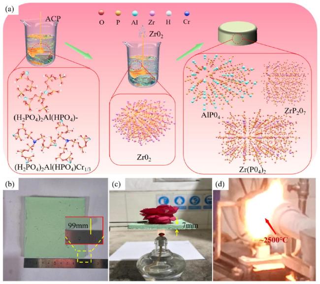
Fig. 1. (a)Reaction flow chart of aluminum chromium phosphate solution with nano- ZrO2 for the preparation of zirconium-based phosphate ceramics (b)Optical photographs of large-sized samples (c)superior thermal insulation (d) ablation resistance. |
3.2. Microstructural characterization
The macroscopic morphology of the zirconium-based phosphate ceramic material, which exhibits a light green color, is clearly visible in Fig. 2(a), and the XRD data indicate that the main physical phases are ZrO2 as well as zirconium-containing phosphate materials (Fig. 2(b)). The microstructure of the zirconium-based phosphate ceramic material was characterized using SEM. The SEM images in Fig. 2(c) clearly shows that the zirconium-based phosphate ceramic material mainly consists of white and gray-white phases that were identified by EDS analysis to be ZrO2 and zirconium-containing aluminum-chromium phosphate. The ZrO2 particles are densely surrounded by fine zirco-nium-containing phosphate particles (Fig. 2(d)). These particles are formed by crosslinking polycondensation to form a three-dimensional mesh structure distributed around the ZrO2, thus effectively bonding the larger particles together.
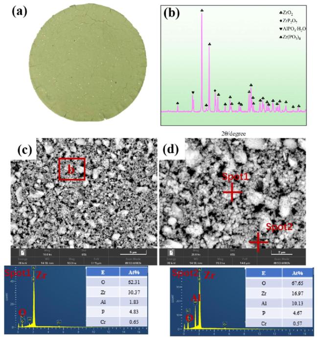
Fig. 2. Structural characterization of zirconium-based phosphate ceramics (a) Optical photograph, (b) XRD pattern, (c) SEM image, (d) Large square view of the marked region in (c). |
To further analyze the microstructure of the zirconium-based phosphate material, this study utilized transmission electron microscopy to analyze the phase distribution. Fig. 3(a) shows a high-angle annular dark-field (HAADF) image of the zirconium-based phosphate material, which shows that the phases are closely combined, and no defects such as cracks and pores appear. Subsequently, EDS mapping was used to analyze the material, as shown in Fig. 3(b). Clearly, the material is divided into two phases. Phase I is primarily composed of zirconia phase and phosphorus phase enriched on its surface, while Phase II is dominated by aluminum phosphate phase. Comparative analysis revealed a distinct selective enrichment of phosphorus in Phase I. The formation mechanism of this phenomenon can be attributed to the following synergistic processes[1-3]: first, Al3+ and Cr3+ preferentially coordinate with phosphate ions in the solution to form an amorphous AlPO4-CrPO4 network structure; second, due to the significant interfacial energy difference between this amorphous network and the zirconia surface, free phosphate ions (PO4 3-) that did not participate in coordination are expelled during the condensationcrystallisation process; Subsequently, these negatively charged phosphate ions are captured by the abundant Zr-OH2 +/Zr-OH groups on the zirconia surface through electrostatic interactions; Finally, during the dehydration-condensation process, the phosphate ions form stable compounds with the zirconia surface through the formation of Zr-O-P covalent bonds or the generation of zirconium pyrophosphate (ZrP2O7, confirmed by XRD in Fig. 2b), thereby creating a characteristic phosphorus enrichment layer on the surface of the zirconia particles. Moreover, it is found that the atomic percentages of oxygen and zirconium were found to be relatively high, reaching 59.22% and 33.74% respectively. The selected-area electron diffraction (SAED) of the zirconium-based phosphate material provides a deeper understanding of the crystal structure of the composite phase. Fig. 3(c) clearly confirms that the two phases are closely combined and that no obvious intermediate phase has formed. The electron diffraction pattern of ZrO2 was analyzed, and the interplanar spacing was measured to be 3Å (Fig. 3(d)), corresponding to the (111) plane. The interplanar spacing of the (-211) plane measured in another direction was 2.78Å. Therefore, this ZrO2 mainly had a cubic phase structure. The corresponding aluminum-containing phosphate phase was mainly amorphous. No lattice fringes appear in this image, and only a small number of diffraction spots exist in the electron diffraction pattern (Fig. 3(f)).
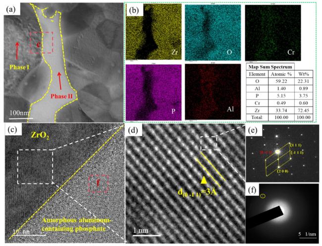
Fig. 3. Microstructural Analysis of Zirconium-Based Phosphate Material. (a) High-Angle Annular Dark-Field (HAADF) Image, (b) EDS Mapping Data of the Area in Figure (a), (c) Selected-Area Electron Diffraction of the Two-Phase Region, (d) Selected-Area Electron Diffraction of Phase I, (e) Electron Diffraction Pattern of Phase I, (f) Electron Diffraction Pattern of Phase II. |
3.3. Thermal stability and mechanical properties of the zirconium-based phosphate ceramic
The dynamic oxidation of zirconium-based phosphate ceramic composites was characterized by conducting TG-DSC tests in an air atmosphere from room temperature to 1400∘C. The TG analysis results in Fig. 4(a) indicate that the sample mass exhibits a significant nonlinear change with increasing temperature, which can be divided into three characteristic transition stages. In the low-temperature range of 0-67∘C, an abnormal increase in mass is observed, primarily due to the inherent hygroscopic properties of phosphate materials. Previous studies have confirmed [1] that such materials can adsorb 2-4wt% moisture at room temperature and form a characteristic "weight gain peak" when heated to ∼70∘C. As the temperature rises to the 67-800∘C range, the sample mass decreases continuously from 102.1% to 97.9%. This mass loss process involves multiple mechanisms [2,3], including the gradual removal of physically adsorbed water and crystalline water, condensation dehydration reactions of interlayer and framework hydroxyl groups, and the release of volatile phosphorus-oxygen species during the condensation or phase transformation of a small amount of phosphate. Notably, in the high-temperature range of 800-1400∘C, the mass change exhibits a fluctuating pattern of first increasing then decreasing: the slight mass increase observed between 800-1200∘C (97.9%→98.4%) can be attributed to the reoxidation process of lowvalent ZrO2-x and P2O5-x components in the material [4], while the subsequent decrease in mass (98.4%→97.8%) observed in the 1200-1400∘C range is directly related to the high-temperature decomposition reaction of pyrophosphate (ZrP2O7→ZrO2+P2O5↑). This conclusion is highly consistent with the phase evolution results shown in Fig. 4(d) for the 1200-1500∘C temperature range. The aforementioned mass change characteristics systematically reveal the complex thermal transformation behavior of phosphate materials at different temperature intervals.
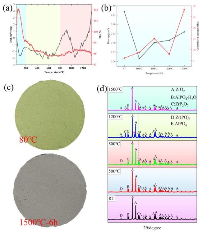
Fig. 4. (a) TG-DSC data of zirconium-based phosphate ceramic, (b)Density and compressive strength data after treatment at different temperatures, (c)Optical photographs before and after treatment at 1500∘C, (d) XRD patterns after treatment at different temperatures. |
In order to analyze in more detail, the heat resistance and the changes in the morphology of the phases during the warming process, a long-time sintering test was performed in a muffle furnace. Fig. 4(b) presents the changes in density and compressive strength after treatments at different temperatures, wherein the density first decreases and then increases. By contrast, the compressive strength initially increases gradually with the temperature rise, then gradually decreases, and finally increases again. This indicates that the temperature treatment affects the fundamental properties of the material. Therefore, the microstructural changes of the phases under different temperature treatments must be examined.
To that end, Fig. 4(c) shows macroscopic photographs of the samples after curing at room temperature and after treatment at 1500∘C for 5 h. The samples before and after treatment are intact and crack-free, and the shrinkage rate is almost zero, verifying the excellent heat resistance. The differences in the changes after different temperature treatments can also be seen in the XRD patterns of the prepared zir-conium-based phosphate ceramics (Fig. 4(d)), which contain more amorphous diffraction peaks after curing at room temperature. Further, crystallization occurs with the gradual loss of physical phases such as bound water as the temperature rises. The complete disappearance of the diffraction peaks of AlPO4H2O after treatment at 800∘C is attributed to the dehydration of AlPO4H2O into AlPO4. The disappearance of the diffraction peaks of ZrP2O7 from 1200∘C to 1500∘C is mainly due to the transformation of ZrP2O7 into ZrO2, and after a long time of treatment at 1500∘C, the samples gradually form phosphate materials with ZrO2 as the main physical phase.
Fig. 5 shows microstructural maps of the surface and cross-sections of the zirconium-based phosphate ceramic material after treatment at room temperature, 500∘C,800∘C,1200∘C, and 1500∘C. The zirconiumbased phosphate ceramic material exhibits a denser microstructure both on the surface and in the cross-section after curing at room temperature. With increasing temperature, the internal microstructure of the sample after treatment at 500∘C starts to decrease in densification, which is manifested by the appearance of loose pores in the internal microstructure (Fig. 5(b)). This characteristic is due to the heat absorption and volatilization of the residual free water sequestered in the sample by the ambient temperature curing. This micromorphological transformation phenomenon is consistent with the density change before and after the treatment temperature (Fig. 4(b)), where the density decreases from 2.469 g/cm3 at room temperature to 2.216 g/cm3 at 500∘C. As the treatment temperature continues to increase, the particles on the sample surface and in the cross-sectionm, as well as the particle boundaries, all become progressively more pronounced, and a similar phenomenon of sinter crystallization occurs. When the temperature increases to 1500∘C, white and grayish-white particulate matter on the nanoscale appears on the surface and in the cross-section of the sample, and the particles are distributed in the sample with a close inter-particle arrangement (Fig. 5(d)). Combining these results with the XRD data (Fig. 4(d)) indicates that the white particulate matter is mainly ZrO2 and that the gray phase is primarily AlPO4.AlPO4 mainly plays the role of bonding ZrO2 particles, which also ensures that the zirconium-based phosphate ceramic composites have good high-temperature stability and temperature resistance.

Fig. 5. Surface and cross-section SEM after different temperature treatments (a) Surface SEM after 80∘C curing, (a1) Cross-section SEM after 80∘C treatment, (a2) Magnification at the markers in (a1), (b) Surface SEM after 500∘C curing, (b1) Cross-section SEM after 500∘C treatment, (b2) Magnification at the markers in (b1), (c) Surface SEM after 800∘C curing, (c1) Cross-section SEM after 800∘C treatment, (c2) Magnification at the markers in (c1), (d) Surface SEM after curing at 1200∘C, (d1) Cross-section SEM after treatment at 1200∘C, (d2) Enlargement at the markers in (d1), (e) Surface SEM after curing at 1500∘C, (e1) Cross-section SEM after treatment at 1500∘C, (e2) Enlargement at the markers in (e1). |
Next, based on the systematic microstructural and phase analysis above, the trends in the density and compressive strength data shown in Fig. 4(b) were analyzed, as discussed below.
With the increasing processing temperature, the density is low at first and then becomes higher. The physical phase and morphology analysis above revealed that with low-temperature curing, zirconium-based phosphate ceramic solidifies more free water and part of the bonded water and other phases, increasing the sample mass. This phenomenon is due to the high density and ambient curing. The same low-temperature-cured sample has many defects, which leads to the compressive strength of this sample being the lowest. As the treatment temperature increases, the free and bound water volatilize, and the sample mass and density decrease. When the temperature is 800∘C, the density appears to increase, but the quality decreases. The data in Fig. 4(b) demonstrate that the density augmentation originates mainly from the dominant sample contraction mechanism. This increase in density with increasing temperature is mainly due to the gradual sintering and ceramization of the sample, whereas an increase in the sample mass results in a slow increase in density.
3.4. Thermal insulation properties of zirconium-based phosphate ceramics
In addition, the zirconium-based phosphate ceramic composites exhibit excellent thermal insulation properties. The back temperature of the composite with a thickness of 10 mm was always lower than 193∘C when heated on a high-temperature platform at 400∘C for a continuous period of 30 min. As can be seen from the infrared thermograms in Fig. 6, the temperature difference between the heated side and the back side of the composite sample is stably controlled above 207∘C. Similarly, the thermal conductivity of the material was evaluated using three-dimensional temperature distribution maps during the heating process, as shown in Fig. 6(f-k). Throughout the continuous heating process, a distinct temperature gradient persisted within the sample. The purple box in the figure designates the sample area. The red region at the top represents the temperature at the bottom of the sample, while the opposite side of the box corresponds to the top. By drawing a line from the top of the sample to the temperature axis, the temperature at the top can be observed to increase with the heating time. However, the rate of temperature rise is slow, and the variation in the width of the isotherms is minimal. This indicates that the sample has low thermal conductivity at this temperature, demonstrating excellent insulating properties. The thermal conductivity in the Z-direction after different temperature treatments is 1.027,0.827,0.934,1.221, and 1.794 W/m⋅K (The data are presented as shown in S1). The thermal conductivity decreases and then increases as the temperature increases, which illustrates that the zirconium-based phosphate ceramic materials should be treated at certain temperatures. The changes in the combination of the mixture phases, microscopic morphology, and density indicate that the thermal conductivity of the zirconium-based phosphate ceramic materials decreases due to the formation of a certain pore structure in the bulk of the heat-treated sample. However, the phosphate phase gradually transforms and volatilizes at an excessively high temperature, and ceramization occurs, which increases the thermal conductivity.
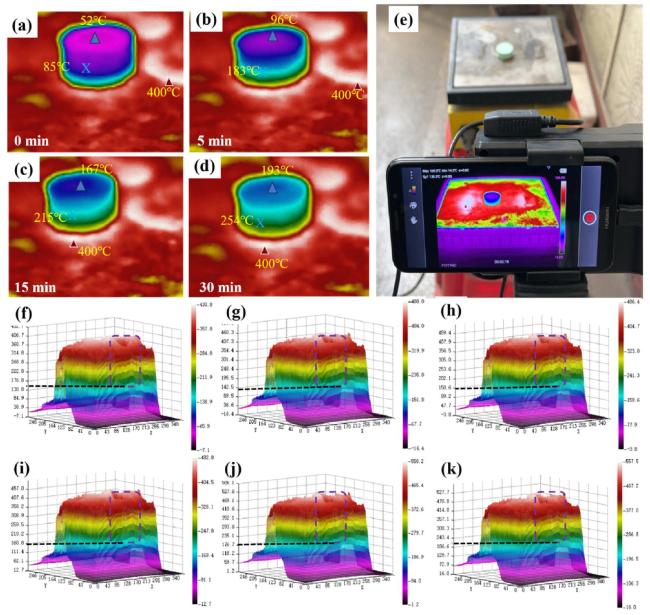
Fig. 6. Infrared thermography of platform heating of zirconium-based phosphate ceramics. (a-d) Infrared thermography of the sample, (e) Optical photos of the experimental process, (f-k) Three-dimensional temperature distribution data. |
A higher temperature gun was used to characterize the thermal insulation properties of the samples. Fig. 7 shows the infrared thermograms of a 10-mm-thick zirconium-based phosphate ceramic material under a high-temperature gun. After achieving a surface temperature of 727∘C with 240 s of continuous heating, the back temperature reaches a maximum of 156∘C. Thus, cooling of 571∘C in the thickness direction is achieved. Moreover, an obvious temperature gradient curve also appears at the heating surface in Fig. 7(b)(c), indicating that the zirco-nium-based phosphate ceramic material has excellent thermal insulation properties in both the Y- and Z-directions. Similarly, Fig. 7(e) depicts the three-dimensional temperature distribution of the sample during ablation by a butane torch. Analysis of the overall isothermal curves reveals both longitudinal and transverse temperature gradients across the sample. Notably, the width of the isothermal lines gradually increases from the high-temperature regions to the low-temperature ones, which suggests that high-temperature zones are localized. As a result, heat accumulates at the ablation site and cannot rapidly disperse throughout the sample. These findings further confirm the outstanding insulating properties of the sample.
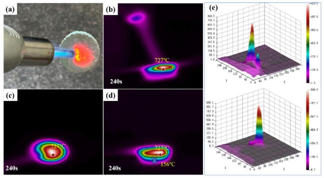
Fig. 7. Infrared thermography of zirconium-based phosphate ceramic material heated by butane torch for 240 s, (a)Optical photos of the experimental process, (b-d) Infrared thermography of the sample, (e)Three-dimensional temperature distribution data. |
The insulation mechanism was also analyzed and is schematically illustrated in Fig. 8. The thermal conductivity of the zirconium-based phosphate ceramics is mainly composed of the gas thermal conductivity (λg), solid thermal conductivity (λs), radiation (λr), and convection (λ con). In this case, λ con is negligible because the pore size of the homogeneous composite is much less than 1 mm. The mean free path of gas molecules is given by Eq. (3). In this equation, λg represents the mean free path of air molecules, k is the Boltzmann constant, T is the thermodynamic temperature, d is the effective diameter of the molecules, and p is the gas pressure. As can be seen from the formula, the mean free path of gas molecules is positively correlated with temperature. When the temperature rises, the mean free path increases significantly to approximately 500 nm, which is larger than the majority of internal pores of the zirconium-based phosphate material (the pore size distributions after various heat treatments are shown in Fig. S2). Thus, the mean free path can be neglected [36,37]. During heating, thermal energy is transferred mainly through the phosphate phase and its interfaces. The phonon scattering effect dissipates a significant amount of energy as it passes through the abundant interfaces of the matrix.
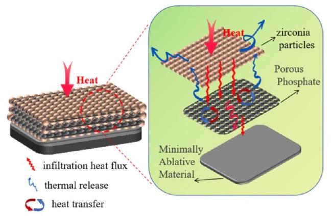
Fig. 8. Thermal insulation mechanism of zirconium-based phosphate ceramic materials. |
Consequently, the composite exhibits excellent thermal insulation properties. The effective synergy of low thermal conductivity and high mechanical properties makes these zirconium-based phosphate ceramics among the most competitive candidates for thermal protection in extreme aerodynamic thermal environments.
3.5. Ablation performance
Next, the zirconium-based phosphate ceramic was tested at 2527∘C under ablation with an oxyacetylene flame for 30 s. During ablation, the temperature of the ablated surface of each sample was tested using an infrared thermometer, and the back temperature of each sample was measured using a thermocouple. The temperature of the ablation surface rapidly reached 2527∘C, whereas the back temperature of the sample peaked at 247∘C (Fig. 9(a)). Thus, the sample cooled down by at least 2280∘C over a distance of 10 mm in the longitudinal direction, confirming its excellent thermal insulation properties. Fig. 9(b) shows the microscopic topography of the ablated surface as a whole, demonstrating that many micrometer-sized holes had formed on the surface; the surface profile after ablation is shown in Fig. S3. The intricate mechanism governing the generation of this extensive array of micropores on the ablated surface is elucidated in Fig. 9(c). Under elevated temperatures, phosphates melt, during which the molten phosphates encapsulate a profusion of zirconia particles. As ablation proceeds, the molten phosphates and internal components partially volatilize and decompose, creating voids. Simultaneously, zirconia particles initiate a process of sintering-induced growth. Ultimately, a distinct hierarchical structure is formed, characterized by a zirconia layer interspersed with a pore-structured layer derived from the residues of volatile decomposition. Thermodynamically, the volatilization and decomposition processes play a crucial role in dissipating substantial amounts of heat. By efficiently removing thermal energy, these processes impede the inward conduction of heat, thus endowing the material with outstanding high-temperature insulation capabilities.
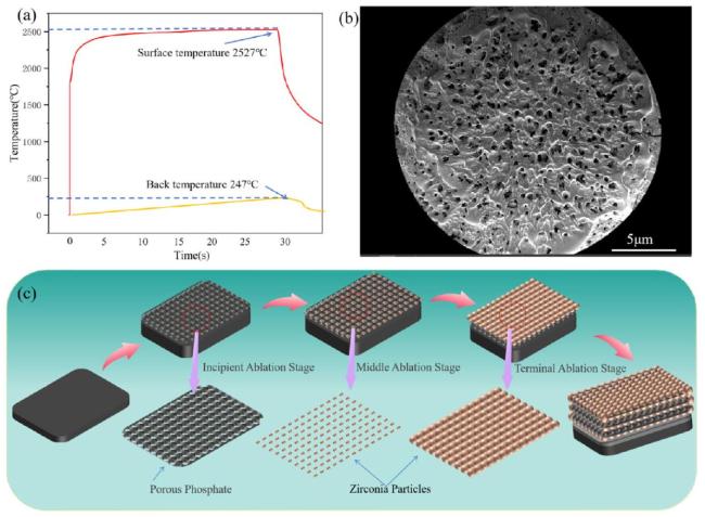
Fig. 9. (a)Ablation surface and backside temperature profiles, (b)Overall micro-morphology of the surface after ablation, (c)Mechanistic Diagram of Micropore Formation on Ablated Surfaces. |
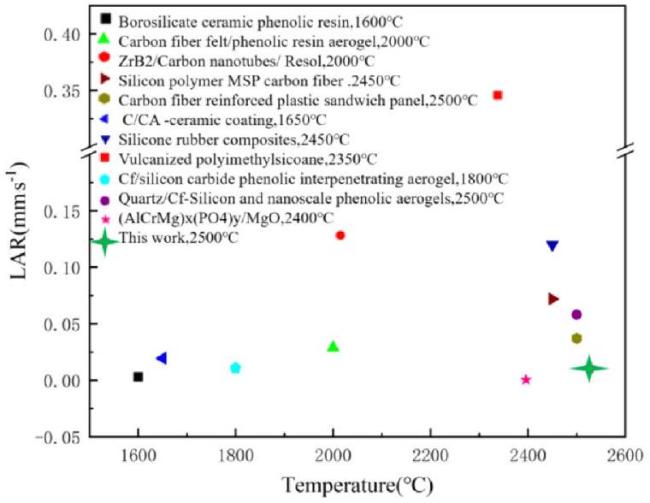
Under oxyacetylene flame ablation at 2500∘C, the zirconium-based phosphate material exhibited mass ablation and linear ablation rates of 0.0173 g/s and 0.0114 mm/s, respectively. In this case, mass ablation primarily resulted from three concurrent mechanisms: (i) thermal volatilization, (ii) pyrolytic decomposition, and (iii) mechanical erosion. During high-temperature ablation, the phosphate phase underwent progressive decomposition and molten-phase migration, while the exposed ZrO2 ceramic phase on the ablated surface experienced grain coarsening through sintering. The ZrO2-rich layer that formed in situ contributed significantly to the ablation resistance of the material through two key effects: (1) the formation of a protective ceramic barrier, and (2) enhanced thermal stability via sinter-induced densification. Fig. 10 comparatively evaluates the linear ablation rates of zirconium-phosphate-based materials and other refractory materials under extreme thermal conditions. The experimental data reveal that the zirconium phosphate system demonstrates a 15-30% lower linear ablation rate than conventional ultra-high-temperature ceramics while maintaining comparable structural stability above 2500∘C. Although its linear ablation rate is slightly higher than that of some polymer-derived ceramics and aerogels, the zirconium phosphate exhibits a substantially higher operational temperature threshold, exceeding these organic-inorganic hybrids by 700-900∘C. This unique combination of exceptional ablation resistance (0.0114 mm/s at 2500∘C) and exceptional thermal stability (up to 2500∘C) positions this zirconium phosphate material as a promising candidate for thermal protection systems requiring balanced performance under extreme thermomechanical loads.
3.6. Ablation mechanism
Fig. 11 shows the microscopic morphology and composition of the sample ablation surface after the sample was ablated in the oxyacetylene flame for 30 s. The ablated surface has obviously formed a dense white layer of material, and combining this finding with the EDS results (Fig. 11(d)(e)) indicates that the white material phase is ZrO2. The ZrO2 oxide layer formed in the central zone of ablation is much denser, with no obvious defects. The ablation transition zone forms a massive ZrO2 of approximately 20μ m, whereas the ablation transition zone forms smaller ZrO2 clasts. The reason for the formation of this unique shape is that when the oxyacetylene flame reaches the surface of the sample, the ablation center area preferentially undergoes high-temperature ablation. During which the phosphate phase decomposes and melts at high temperature, and the ZrO2 phase leaks out of the surface. This ZrO2 then grows with the help of the molten aluminum phosphate and heat, flat sintering to a certain degree, which is the most important factor in the formation of this unique shape. Thermal ablation occurs later in the ablation transition and edge zones than it does in the ablation center. Further, the ablation flame has no mechanical impact, the phosphate phase melts and sinks, and ZrO2 in the edge region grows slowly, resulting in the appearance of a certain gap. Ablation occurs on the surface as the decomposing phosphate phase both volatilizes and melts, and the molten phosphate phase deposits on the lower layer. Hence, the phosphate phase is not detected on the surface; instead, a large amount of bare ZrO2 leakage occurs during ablation, and the molten phosphate phase acts as a binder to firmly bind the surface layer of ZrO2 during ablation, thus improving its resistance to ablative corrosion.

Fig. 11. Microscopic morphology of the ablated surface of zirconium-based phosphate ceramics. (a) (a1) Ablation central area. (b) (b1) Ablation transition zone. (c) (c1) Ablation edge zone. (d)Mapping of the central ablation area. (e)Elemental spectrum and atomic percentage (weight percentage) from mapping data. |
The microstructure of the sample cross-section after ablation is shown in Fig. 12. The ablation layer has a dense ZrO2 layer with a thickness of approximately 2.12 mm (Fig. 12(a)), and the oxide layer is structurally intact without cracks or other defects, which can effectively prevent the ablation flame from directly destroying the interior of the sample. The structural changes can be clearly seen in the ablation transition layer in Fig. 12(b), where the top-down microphase gradually becomes less dense, and ZrO2 tends to increase in granularity, with no obvious structural deformation or visible collapse. This is attributed to the ablation that occurs in this region, where heat transfer from the surface layer to the transition ablation layer results in the decomposition of the phosphate in this layer. This decrease is also caused by the release of pyrolysis gases, raising the pressure inside the sample, which attenuates the continued transfer of heat to the lower layers to some degree (Fig. 12(d)). On the other hand, the sintering growth of ZrO2 particles is hindered by the pressure, which gradually forms 2-6μ m particles with smooth surfaces and a certain pore structure between the particles. The heat from the ablation surface is transferred to the original material layer shown in Fig. 12(c), but by then, its heat has already been mostly dissipated. Hence, the bottom of the layer is at a maximum temperature of 247∘C, which is generally low, and the bonding morphology features within the zirconium-based phosphate ceramic are fully preserved.
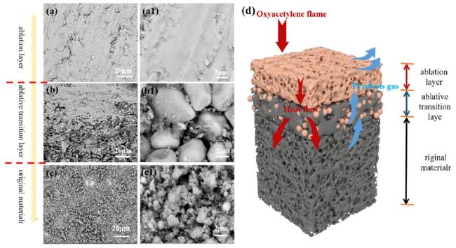
Fig. 12. Micro-morphology of zirconium-based phosphate ceramics in cross-section after ablation. (a)(a1) Ablation layer. (b) (b1) Ablation transition layer. (c) (c1) Original material layer. (d) Ablation mechanism diagram. |
This comprehensive analysis of the ablation process reveals the underlying mechanism, as illustrated in Fig. 12(d). The post-ablated sample exhibits a distinct gradient structure comprising three well-defined layers from top to bottom: (i) the ablation layer, (ii) the ablation transition layer, and (iii) the original material layer. During the initial ablation stage, the surface of the ablation layer experiences severe degradation, including the thermal decomposition and melting of phosphate phases, accompanied by the sintering-induced grain growth of zirconia particles. As ablation progresses to the intermediate stage, the accelerated sintering of zirconia particles forms a continuous zirconiarich protective layer (ablation layer), which effectively impedes flame penetration. Concurrently, the molten phosphate phase partially volatilizes and partially migrates downward, with the descending molten phosphate providing thermal energy to promote gas evolution in the transition layer. In the final ablation stage, the rapid release of pyrolysis gases from the transition layer generates significant internal pressure, which establishes a thermal barrier that effectively limits heat transfer to deeper regions. This mechanism maintains the temperature of the bottommost layer at 247∘C, thereby preserving the structural integrity of the original material. The resulting multi-layered architecture demonstrates synergistic protection: the ZrO2-rich surface layer provides ablation resistance, and the porous transition layer offers thermal insulation to protect the original material layer below.
The above results show that zirconium-based phosphate ceramics undergo complex oxidation, decomposition, volatilization, and other processes at high temperatures. The relevant literature states that the possible reactions of zirconium-based phosphate ceramics at high temperatures are as follows [47-49]:
${\mathrm{C}\mathrm{r}\mathrm{O}}_{3\left(\mathrm{l}\right)}\to {\mathrm{C}\mathrm{r}\mathrm{O}}_{\left(\mathrm{g}\right)}+{\mathrm{O}}_{2(g)}$
${\mathrm{C}\mathrm{r}}_{2}{\mathrm{O}}_{3\left(\mathrm{l}\right)}\to {\mathrm{C}\mathrm{r}\mathrm{O}}_{\left(\mathrm{g}\right)}+{\mathrm{O}}_{2(g)}$
${\mathrm{P}}_{2}{\mathrm{O}}_{5\left(\mathrm{l}\right)}\to {\mathrm{P}}_{2\left(\text{ }\mathrm{g}\right)}+{\mathrm{O}}_{2(g)}$
${\mathrm{Z}\mathrm{r}\mathrm{P}}_{\left(\mathrm{l}\right)}\to {\mathrm{Z}\mathrm{r}\mathrm{P}}_{(g)}$
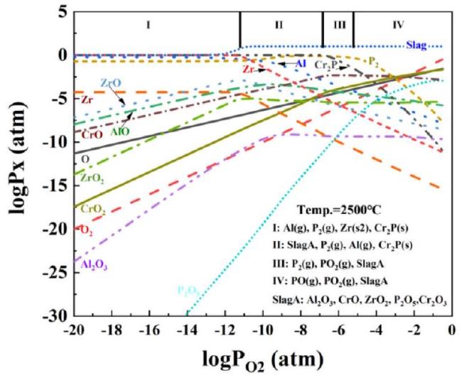
In order to understand the ablation behavior of the zirconium-based phosphate ceramics further, the equilibrium thermodynamics of these materials was calculated, and the gas volatility was plotted as a function of PO2 and temperature. The graph was obtained using FactSage 7.3 software and data from the NIST-JANAF database, and the fluctuation graph corresponding to 2500∘C in Fig. 13 shows that the wave diagrams of the zirconium-based phosphate ceramic materials as a function of the PO2 partial pressure can be divided into four states[50]:
Fig. 13. Fluctuation diagram of zirconium-based phosphate ceramics at 2500∘C. |
(I) When PO2<10-11.5, the oxygen potential is low, the oxides are unstable, and only gas-phase monomers exist, including aluminum, zirconium, dichromated phosphorus, and phosphorus, with no condensed phase.
(II) When 10-11.5<PO2<10-7, elemental gas phases such as phosphorus, aluminum, and di-zirconiated phosphorus are still present; the oxide phase is unstable; and the condensed phases are Al2O3,CrO, ZrO2,P2O5, and Cr2O3.
(III) When 10-7<PO2<10-5, PO2 gas appears, the gas phase of phosphorus decreases, and the condensed phase consists of Al2O3,CrO, ZrO2,P2O5, and Cr2O3 slag.
(IV) When PO2>10-5, the phosphorus gas phase is completely transformed into PO and PO 2 gas phases, and the condensed phases are Al2O3,CrO,ZrO2,P2O5, and Cr2O3.
In conclusion, the volatilization of low-melting-point phases occurs first in the zirconium-based phosphate ceramics under ablation at 2500∘C. As the partial pressure of oxygen decreases, Al-O and Cr-O gas phases begin to appear. Finally, the Zr-O gas phase appears, suggesting that ZrO2 is the most difficult phase to volatilize because it has the lowest vapor and decomposition pressures. The formation of a dense zirconia layer on the surface after ablation was also verified.
4. Conclusion
Zirconium-based phosphate ceramics with ablation resistance and thermal insulation properties were prepared by crosslinking and polycondensation curing of aluminum-chromium phosphate and ZrO2. The zirconium-based phosphate ceramics exhibited excellent thermal stability (Fig. 4(a)), high mechanical strength (Fig. 4(b)), low thermal conductivity (Figs. 6 and 7), and excellent high-temperature thermal insulation properties (Fig. 9(a)). The composites were evaluated by oxyacetylene ablation at 2527∘C for 30 s, and the line and mass ablation rates reached 0.0173 g/s and 0.0114 mm/s, respectively. Due to the formation of a pronounced longitudinal gradient under high-temperature ablation (Fig. 12), a surface temperature of 2500∘C and a maximum back temperature of 247∘C were obtained for a 10 mm longitudinal sample. Thus, the zirconium-based phosphate ceramic exhibited excellent thermal insulation properties at high ablation temperatures. Zirconium-based phosphate ceramics have obvious performance advantages over the traditional integrated heat-insulation materials. Hence, this study indicates a promising new direction for research on ultra-high-temperature thermal protection materials.